Fast-Green Staining of Collagen 3D Network
A revolutionary method for imaging collagen 3D network architecture in large samples with unmatched high-resolution
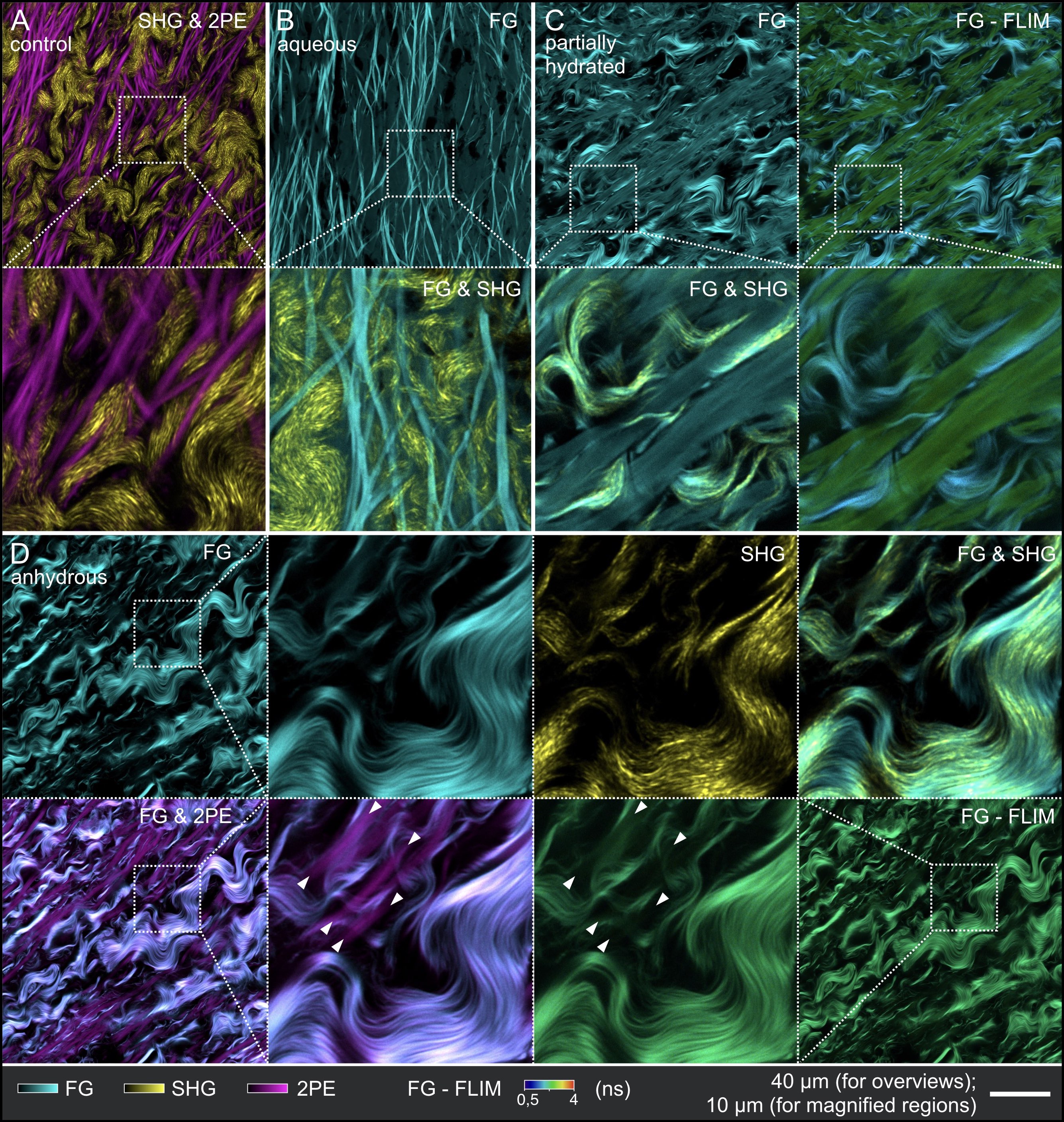
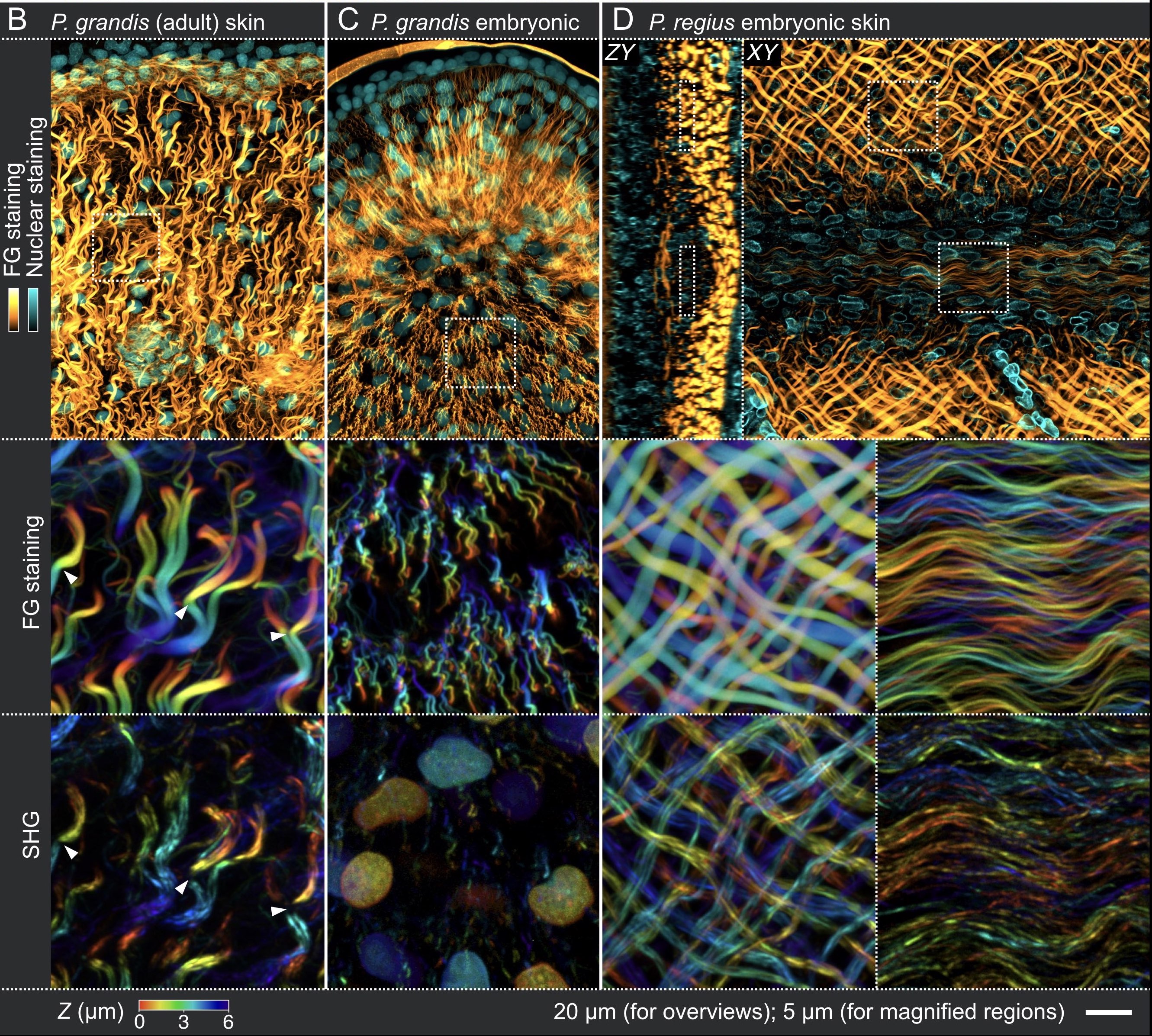
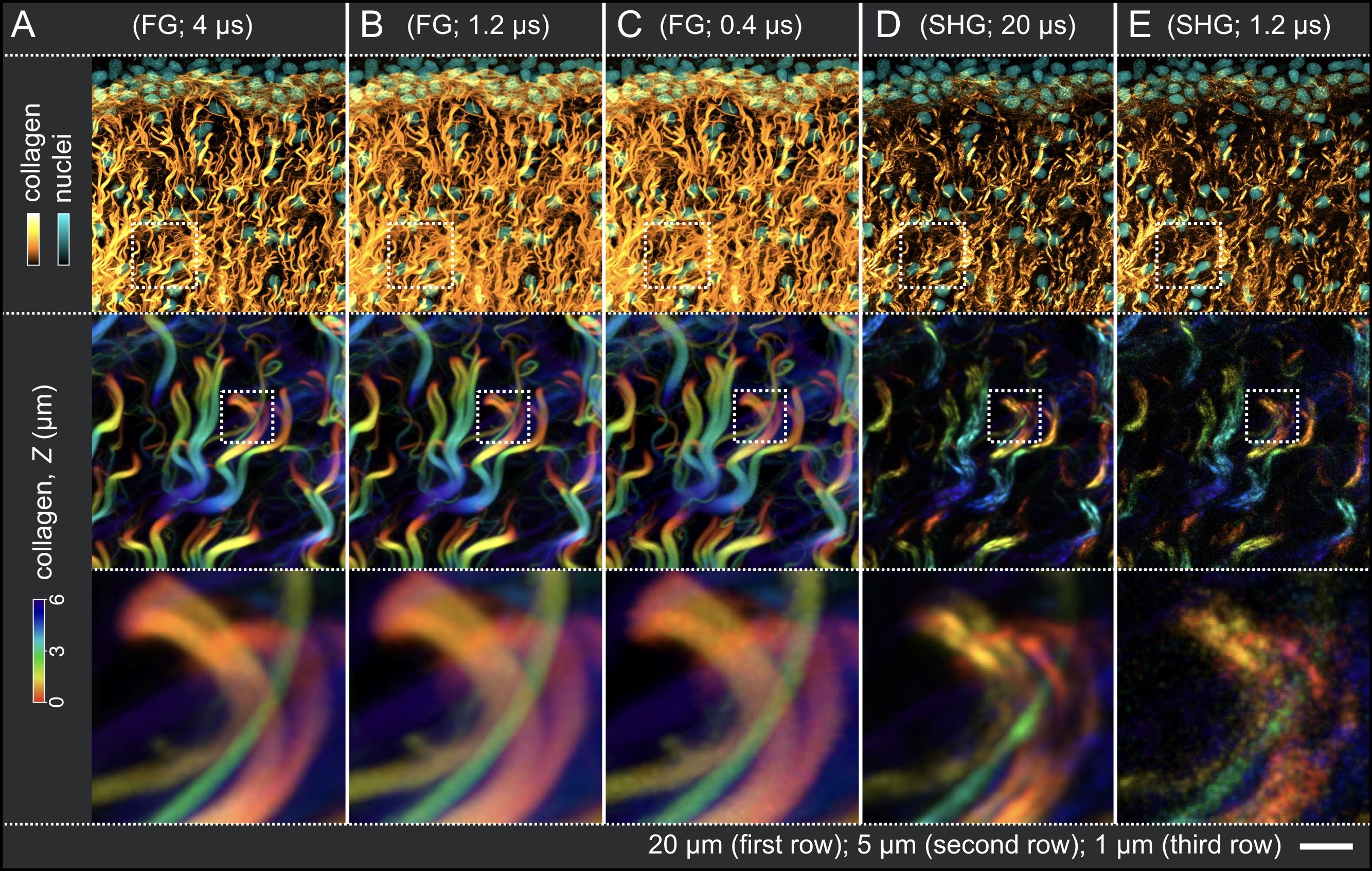
Although notoriously difficult, imaging collagen network architecture, a key element affecting tissue mechanical properties, is of paramount importance in developmental and cancer biology. Here, we introduce a simple and robust method of whole-mount collagen staining with the ‘Fast Green’ dye that provides visualisation of collagen 3D network architecture, via confocal or light-sheet microscopy, with unmatched resolution. This new approach is compatible with tissue clearing techniques and immunostaining.
Our report is available in the following article
High-Resolution Imaging of Collagen 3D Network Architecture in Very Large Samples
Timin Grigorii & Michel C. Milinkovitch
iSciences 26, 106452 (2023)
Correspondence: Michel C. Milinkovitch
Download full-resolution images
Use the photo gallery below to download the full-resolution images used in the various panels of Figure 1, Figure 2, Figure 3, Figure 4 and Figure 5.
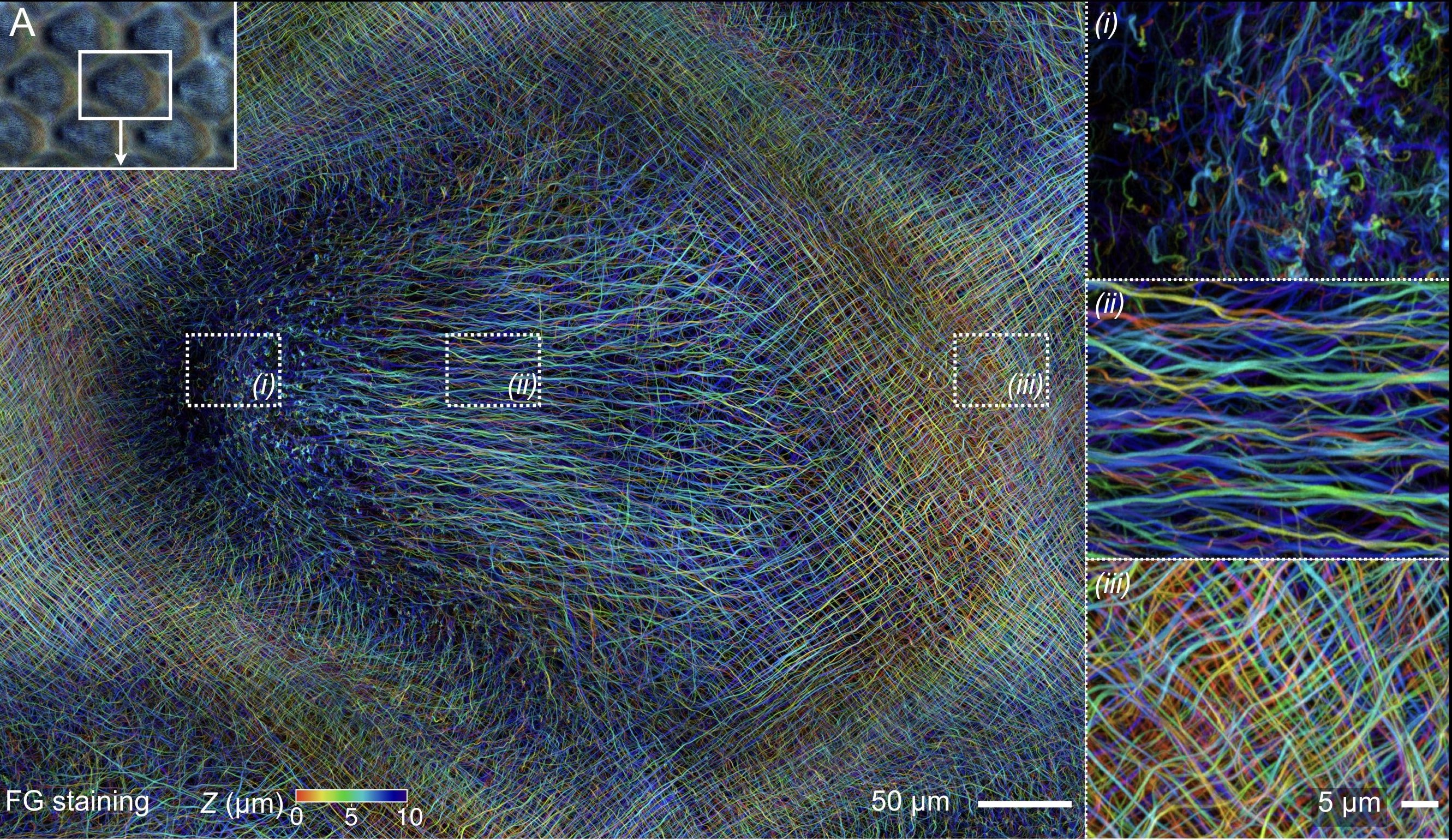
Figure 1B-D
Download full resolution:
Fig1B_FG,YoPRO1 Fig1B_FG Fig1B_SHG Fig1C_FG,DAPI Fig1C_FG Fig1C_SHG Fig1D_FG,LamB1_XZ Fig1D_FG,LamB1_XY Fig1D_FG Fig1D_SHGFigure 2A-E
Download full resolution:
Fig2A_FG,YoPRO1 Fig2A_FG Fig2B_FG,YoPRO1 Fig2B_FG Fig2C_FG,YoPRO1 Fig2C_FG Fig2D_SHG,YoPRO1 Fig2D_SHG Fig2E_SHG,YoPRO1 Fig2E_SHG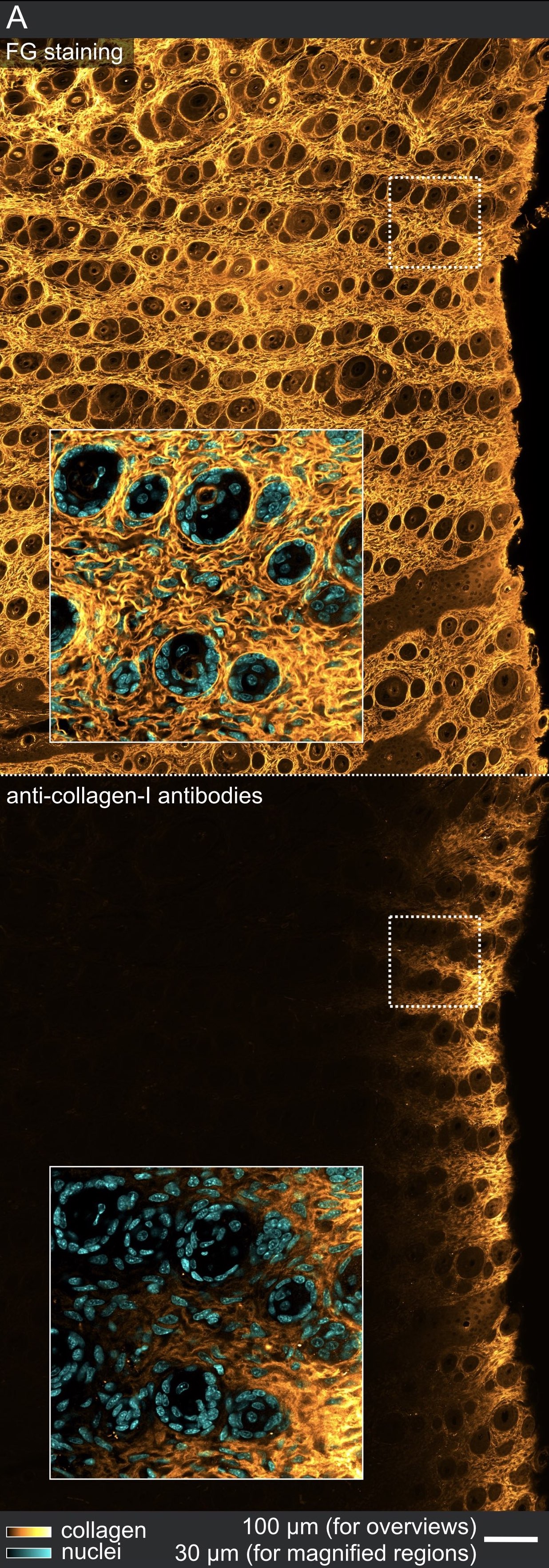
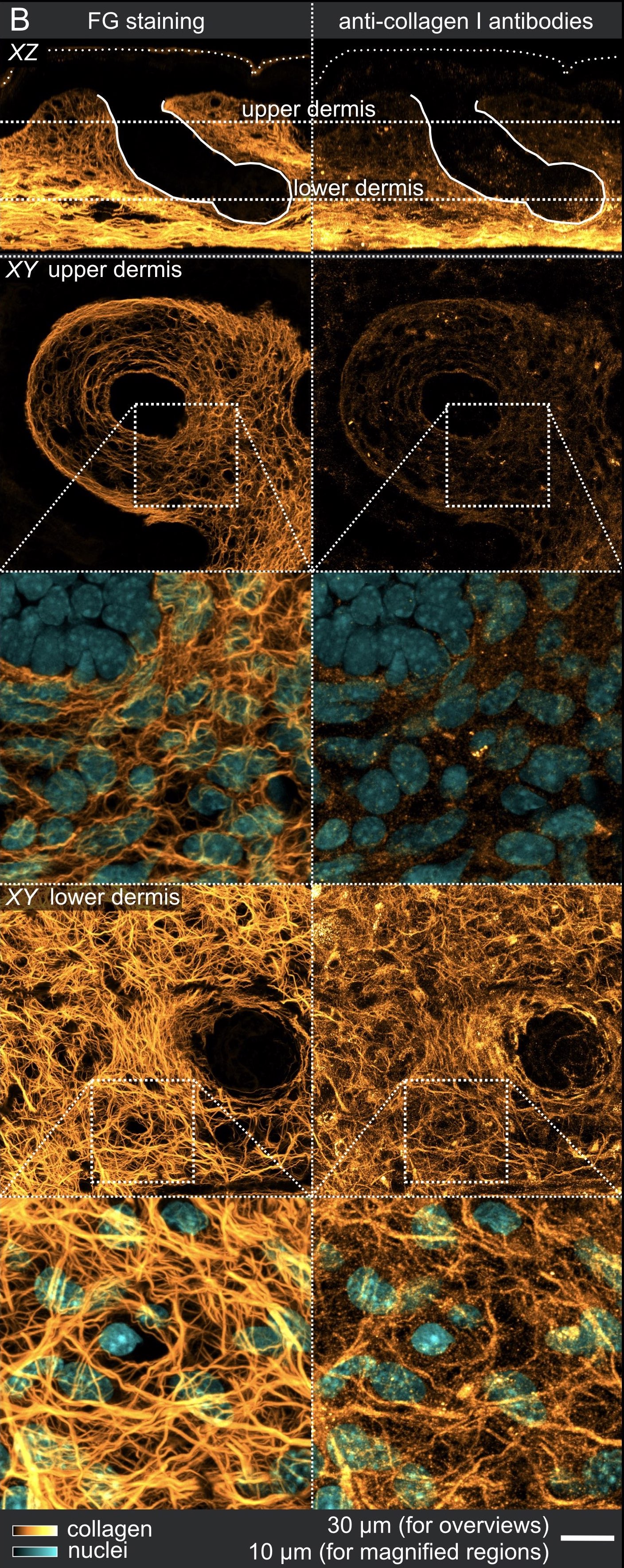
Figure 3B
Download full resolution:
Fig3B_XZ_FG Fig3B_XZ_immunostaining Fig3B_upper_dermis_FG Fig3B_upper_dermis_immunostaining Fig3B_upper_dermis_FG,DAPI Fig3B_upper_dermis_immunostaining,DAPI Fig3B_lower_dermis_FG Fig3B_lower_dermis_immunostaining Fig3B_lower_dermis_FG,DAPI Fig3B_lower_dermis_immunostaining,DAPIFigure 4A
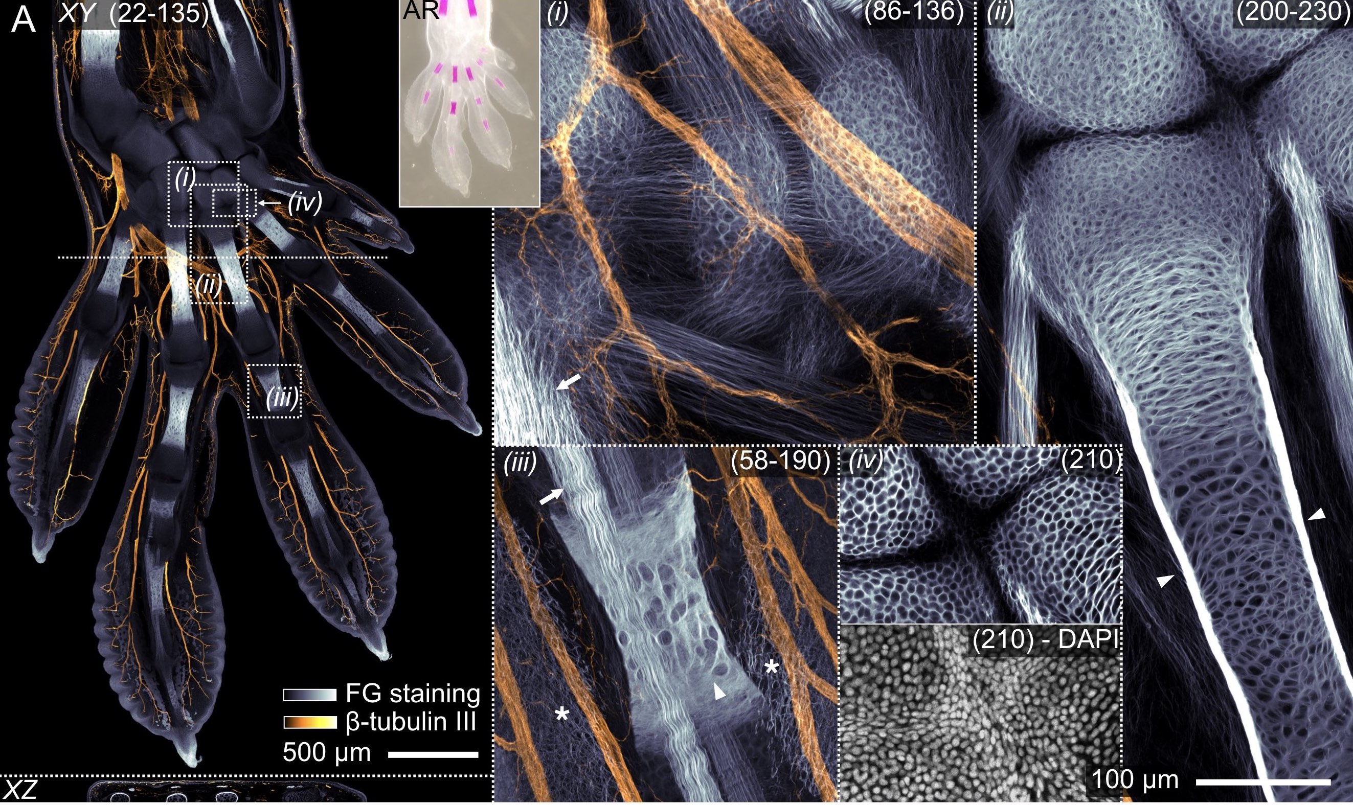
Download full resolution:
Fig4A_overview Fig4A_XZ Fig4A_AR Fig4A_(i) Fig4A_(ii) Fig4A_(iii) Fig4A_(iv)FG Fig4A_(iv)DAPI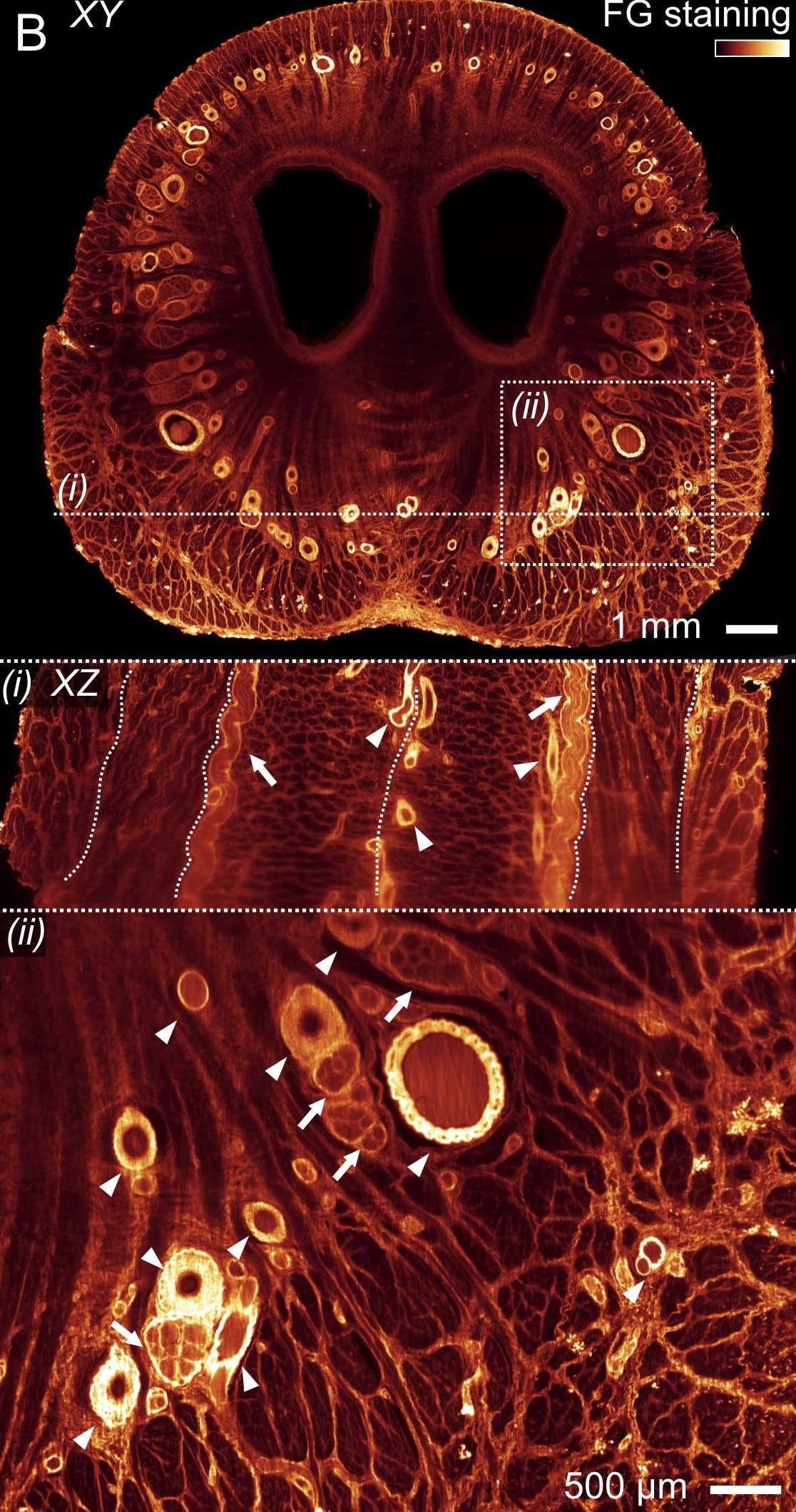
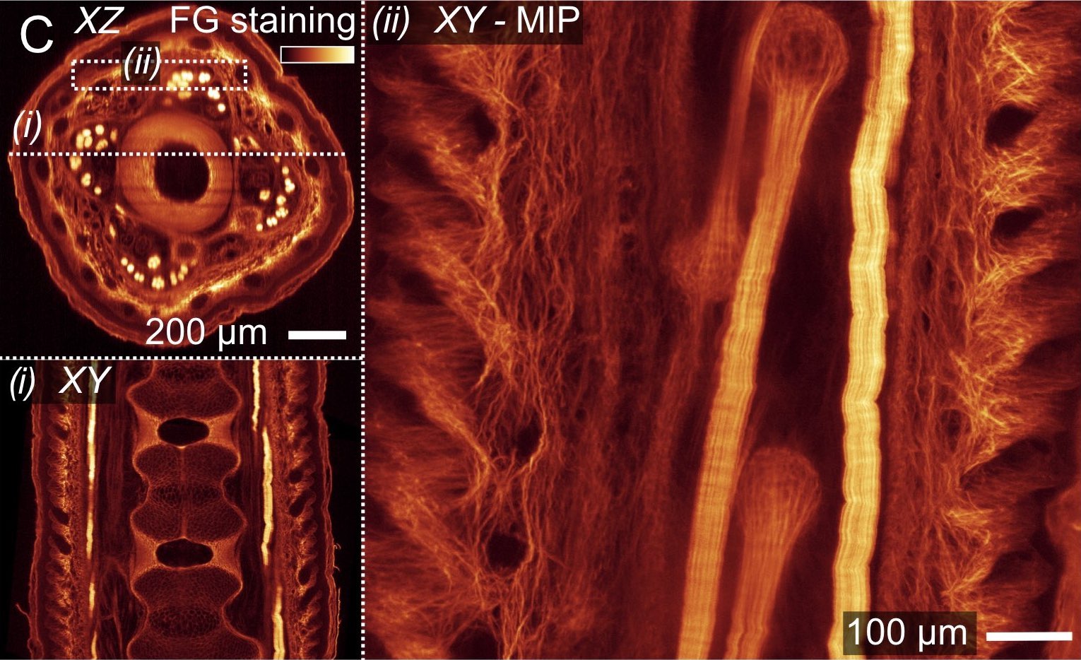
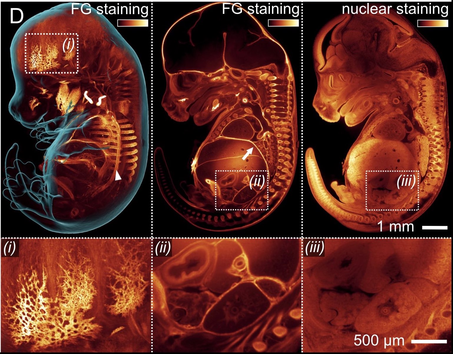
Figure 5